
Patients searching for the best lung cancer specialist in Pune often look for accurate diagnosis, advanced treatment planning, and personalised oncology care tailored to their condition. Lung cancer treatment may involve careful evaluation, timely medical intervention, and structured recovery support designed to improve overall treatment outcomes and patient confidence.
It is a significant health issue worldwide
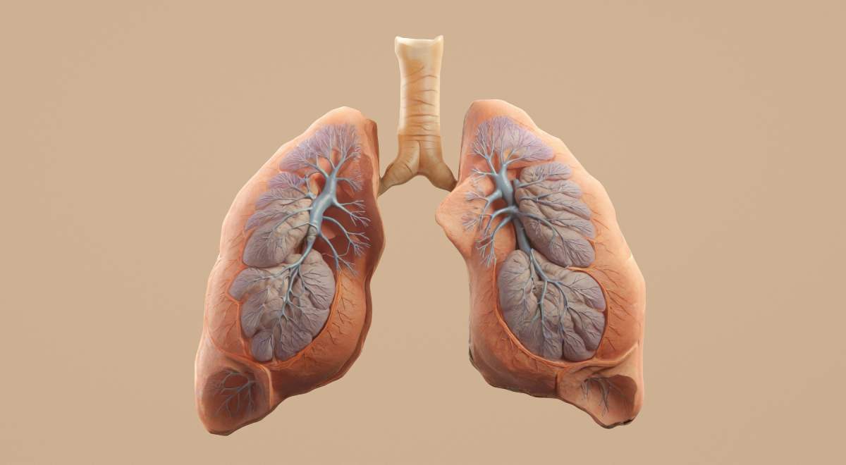
Types Of Lung Cancer
Lung cancer is broadly classified into two main types:
Non-Small Cell Lung Cancer (NSCLC) and Small Cell Lung Cancer (SCLC). NSCLC is the more common type and includes Adenocarcinoma, which is frequently seen in non-smokers and women and usually develops in the outer regions of the lungs; Squamous Cell Carcinoma, which is often associated with smoking and typically arises in the larger airways; and Large Cell Carcinoma, a less common but aggressive form that can occur in different parts of the lungs. Small Cell Lung Cancer (SCLC) is strongly linked to heavy smoking, tends to grow rapidly, and is more likely to spread to other parts of the body at an early stage.
Risk Factors
Early consultation with the best lung cancer specialist in Pune may help patients better understand their condition and available treatment options.


")

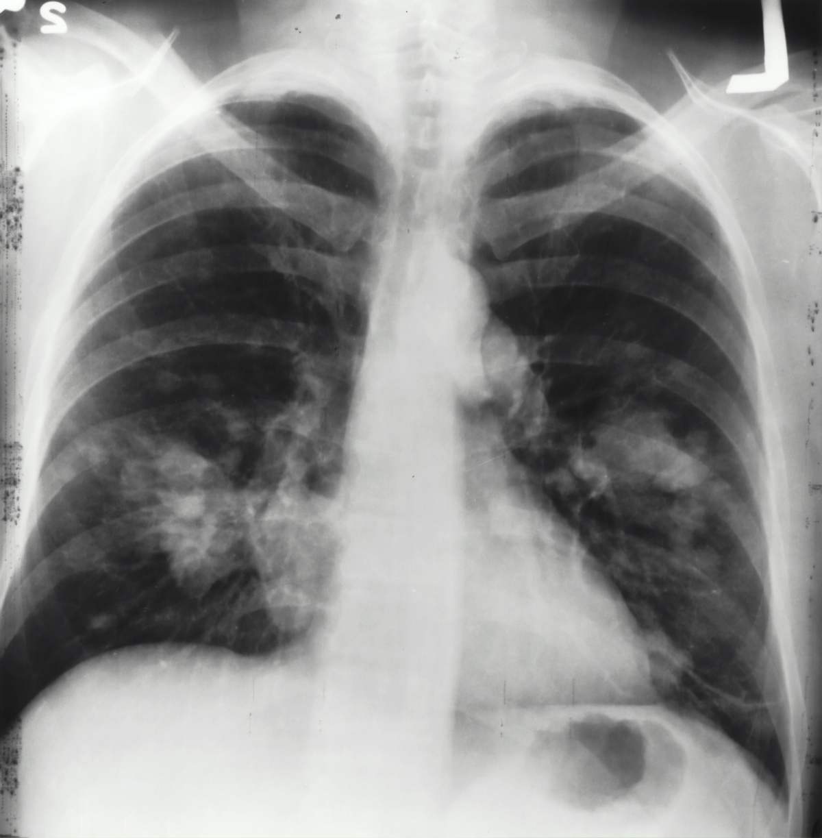
Lung Cancer Diagnosis and Treatment
Patients searching for the best lung cancer specialist in Pune often look for timely diagnosis, advanced treatment planning, and personalised oncology care. Early evaluation of persistent respiratory symptoms can help support accurate diagnosis and treatment decisions.
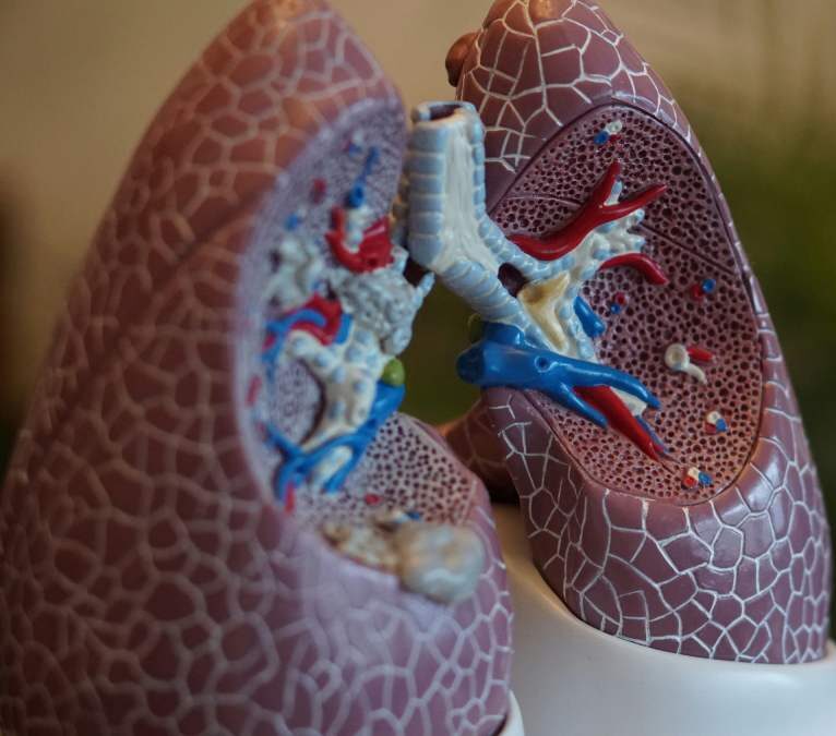
Role of Indocyanine green (ICG) in Thoracoscopic lung surgery
Indocyanine Green (ICG) technology plays an important role in thoracoscopic lung surgery by helping surgeons identify lung segments, blood supply, and tumour boundaries with greater precision during minimally invasive procedures. Patients consulting the best lung cancer specialist in Pune may benefit from advanced surgical techniques such as ICG-guided thoracoscopic surgery for improved surgical accuracy and better recovery support.
Enhanced Visualization
Perfusion Assessment: ICG is used to assess lung perfusion during surgery. After intravenous injection, ICG binds to plasma proteins and emits fluorescence when illuminated with near-infrared light. This helps surgeons visualize the vascular and perfusion status of lung tissues, ensuring adequate blood supply to the remaining lung after resection.
Segmental Resection: For segmentectomy, ICG helps delineate the intersegmental plane. By injecting ICG into the pulmonary artery supplying the segment to be preserved, the boundary between the fluorescent and non-fluorescent areas marks the resection plane, allowing precise and limited removal of lung tissue.
Tumor Localization
Intraoperative Navigation: Preoperative injection of ICG around the tumor can help localize small or deep-seated lesions that are difficult to palpate or visualize during surgery. This technique enhances the accuracy of tumor resection and helps ensure complete removal of the malignancy.
Patients consulting the best lung cancer specialist in Pune are often advised to undergo imaging and biopsy investigations for detailed cancer evaluation.
Identification of Lymph Nodes in Lung Cancer
Accurate identification and assessment of lymph nodes play an important role in determining the stage of lung cancer and planning appropriate treatment. The best lung cancer specialist in Pune may use advanced diagnostic and surgical techniques to evaluate lymph node involvement, helping guide treatment decisions and improve overall patient outcomes.
Sentinel Lymph Node Mapping
Improved Staging
Reduced Morbidity
Advanced technologies such as Indocyanine Green (ICG) fluorescence imaging also help improve surgical precision during thoracoscopic lung surgery by enabling better lymph node identification and more accurate cancer staging. The best lung cancer specialist in Pune may use advanced imaging-guided surgical techniques to support precise treatment planning, targeted procedures, and improved surgical outcomes.
Benefits
Overall, ICG fluorescence imaging represents a significant advancement in thoracoscopic lung surgery and lymph node identification, improving the precision and outcomes of lung cancer surgeries.
Get Expert Care from a Trusted Lung Cancer Specialist in Pune
Choosing the best lung cancer specialist in Pune can make a meaningful difference in treatment planning, recovery support, and overall patient confidence. Early medical consultation may help patients explore suitable treatment options with greater clarity and timely care.