
Introduction
Oral cancer can begin with something that seems easy to ignore, such as a sore that does not heal, a white or red patch, unexplained bleeding, or a lump in the mouth. If these changes persist, getting evaluated by an experienced Oral Cancer Specialist In Pune can help identify the cause and determine whether further testing is necessary. The oral cancer diagnosis process is not based on a single examination. Instead, doctors use a step-by-step approach that may include a detailed medical history, physical examination, biopsy, imaging, laboratory analysis, and staging.
What Is Oral Cancer?
Oral cancer refers to cancer that develops in parts of the oral cavity, including the lips, gums, inner lining of the cheeks, tongue, floor of the mouth, hard palate, and areas behind the wisdom teeth. Cancers occurring farther back in the throat are generally classified separately as oropharyngeal cancers, although symptoms and diagnostic investigations can sometimes overlap.
The disease may initially produce subtle changes. A person might notice a persistent ulcer, thickened area, unusual patch, difficulty moving the tongue, or discomfort while chewing. In some cases, there may be no significant pain in the early stages.
That is one reason early evaluation matters. A suspicious-looking area does not automatically mean cancer. Many mouth lesions are caused by infections, trauma, irritation, dental problems, or other non-cancerous conditions. The purpose of an examination is to find the actual cause rather than assume the worst.
When Should You Get Checked for Oral Cancer?
Knowing when to seek professional advice is an important part of early detection of oral cancer.
You should consider seeing a dentist or doctor if you notice a mouth or throat change that does not improve, particularly when it persists for a few weeks. Warning signs can include:
- A sore in the mouth or on the lip that does not heal
- A white or red patch
- A lump or thickened area inside the mouth
- Persistent mouth pain
- Unexplained bleeding
- Difficulty chewing or swallowing
- Difficulty moving the tongue or jaw
- Persistent numbness in the tongue or mouth
- A lump in the neck
- Changes in speech or voice
- Persistent sore throat
- Unexplained ear pain
- Teeth becoming loose without an obvious dental cause
- Dentures becoming uncomfortable or fitting differently
These symptoms can have many causes and do not necessarily indicate cancer. However, persistent or unexplained changes should be evaluated rather than repeatedly treated at home.
Step 1: Medical History and Risk Assessment
The first part of the oral cancer diagnosis process usually begins with a conversation.
Your doctor may ask when you first noticed the problem, whether it has changed in size or appearance, whether it bleeds, and whether you experience pain or difficulty eating, speaking, or swallowing.
Your medical history also matters. The doctor may ask about
- Tobacco use, including smoking and smokeless tobacco
- Alcohol consumption
- Previous oral or head and neck cancers
- Previous radiation treatment
- Dental or oral problems
- Previous biopsies or suspicious lesions
- Relevant medications
- Previous surgeries
- General health and nutritional status
Tobacco and alcohol are established risk factors for oral cancer, and using them together can increase risk. Human papillomavirus, particularly HPV type 16, is strongly associated with certain oropharyngeal cancers.
This conversation is not about judging lifestyle choices. It gives the doctor useful information for interpreting examination findings and deciding what investigations may be appropriate.
Step 2: Oral and Head and Neck Examination
The physical examination is one of the most important parts of how oral cancer is diagnosed.
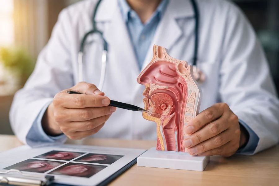
A doctor or dentist carefully examines the mouth, lips, gums, tongue, cheeks, palate, and other visible areas. The neck and face may also be examined for swelling, asymmetry, or enlarged lymph nodes. The examination can involve both looking and gently feeling the tissues.
The clinician may use a gloved finger to assess areas inside the mouth and check whether a suspicious area feels soft, firm, thickened, fixed, or tender. The neck may also be gently palpated to identify enlarged lymph nodes or other abnormalities.
A comprehensive oral examination can often be completed relatively quickly. It is generally straightforward and does not necessarily involve an invasive procedure.
Why Does the Physical Examination Matter?
Visible changes can sometimes provide the first clue that additional investigation is required.
For example, a persistent ulcer may appear different from a typical traumatic ulcer. Similarly, a patch that remains despite removing a possible source of irritation may need closer assessment. However, visual examination alone cannot confirm cancer.
That distinction is important. A doctor may identify a suspicious lesion during oral cancer screening, but the next step depends on its appearance, location, duration, symptoms, and other clinical factors.
Step 3: Identifying Suspicious Changes
Not every mouth ulcer or patch requires a biopsy.
Doctors consider several factors before deciding whether tissue sampling is necessary. These can include:
- How long the lesion has been present
- Whether it is increasing in size
- Whether it bleeds easily
- Whether it is painful or numb
- Whether it has an unusual surface
- Whether the surrounding tissue feels abnormal
- Whether there is an associated neck lump
- Whether the patient has significant risk factors
- Whether the lesion improves after removing a possible source of irritation
Sometimes a doctor may initially recommend observation or treatment for an obvious non-cancerous cause, followed by reassessment.
If the abnormality persists or appears suspicious, a biopsy may be recommended.
Step 4: Additional Examination of the Throat and Neck
Some areas of the mouth and throat cannot be fully evaluated by simply looking inside the mouth. When necessary, an ENT or head and neck specialist may use an instrument called an endoscope. This is a thin viewing device that allows the doctor to inspect areas that are difficult to see directly.
Depending on the symptoms and suspected location, examination may extend to the back of the throat, base of the tongue, voice box, or other areas of the upper airway. Flexible endoscopic examinations can provide a closer view of areas that are difficult to assess using a standard mouth examination.
The exact examination depends on the location of the suspected lesion. Not every patient needs an endoscopy.
Step 5: Biopsy to Confirm Cancer
If there is a suspicious lesion, a biopsy is often the most important diagnostic step.
A biopsy involves removing a small sample of tissue so that it can be examined under a microscope by a pathologist. A biopsy is considered the definitive way to establish whether oral cavity or oropharyngeal cancer is present.
What Happens During an Oral Biopsy?
The procedure depends on the location and size of the abnormal area. For an easily accessible lesion, the doctor may numb the area with a local anaesthetic and remove a small portion of tissue. This is commonly referred to as an incisional biopsy.
If the entire small lesion can be safely removed, an excisional biopsy may sometimes be considered. For deeper lesions, the procedure may need to be performed in an operating room using appropriate anaesthesia.
The tissue is then sent to a pathology laboratory.
Why Is a Biopsy So Important?
A scan can show an abnormal mass. An examination can identify a suspicious patch. However, neither one can independently provide the same level of confirmation as microscopic examination of tissue. The pathologist examines the cells and tissue architecture to determine whether cancer is present and, when applicable, identifies the type and characteristics of the cancer.
This information becomes important when the medical team starts planning treatment.
Step 6: Fine Needle Aspiration for Neck Lumps
Sometimes the first noticeable sign is not a mouth lesion but a lump in the neck. An enlarged lymph node can occur for many reasons, including infections. However, when the clinical findings raise concern about cancer, doctors may recommend fine needle aspiration, commonly called FNA.
During FNA, a thin needle is inserted into the suspicious lump to collect cells or fluid. The sample is then examined in the laboratory. FNA can be particularly useful when doctors need to determine whether an enlarged neck lymph node contains cancer cells. It can also help investigate whether cancer in a lymph node may have originated elsewhere.
The decision to perform FNA depends on the clinical situation and examination findings.
Step 7: Imaging Tests
Once cancer is suspected or confirmed, imaging may be needed to understand the size and location of the disease. Different imaging methods provide different types of information.
CT Scan
A CT scan uses X-rays to create detailed cross-sectional images of the body. For oral cancer, CT imaging can help doctors assess the primary tumour and examine nearby structures. It can also provide information about lymph nodes and areas that may be difficult to evaluate during a physical examination.
MRI
MRI uses magnetic fields and radio waves rather than X-rays. It can provide detailed images of soft tissues and may be particularly useful when doctors need to understand the relationship between a tumour and nearby structures.
PET-CT
A PET-CT combines metabolic information with detailed anatomical imaging. It may be used in selected patients to look for areas of active disease elsewhere in the body, assess suspected spread, or help with staging and treatment planning.
Not every patient needs every imaging test. The choice depends on the suspected location, confirmed diagnosis, stage, symptoms, and treatment plan.
Step 8: Pathology and Understanding the Diagnosis
After a biopsy, the pathology report becomes a central part of the diagnosis. The report can provide information about whether malignant cells are present and what type of cancer has been identified. Additional microscopic or laboratory assessments may sometimes be required depending on the cancer type and clinical circumstances.
This is where the diagnostic process moves beyond the question, “Is this cancer?”
The medical team also needs to understand:
- What type of cancer is it?
- Where did it begin?
- How extensive is it?
- How deeply has it invaded?
- Are lymph nodes involved?
- Are there other areas that need investigation?
- Are additional laboratory tests needed?
These answers help doctors develop an appropriate treatment strategy.
Step 9: Cancer Staging
After cancer has been confirmed, the next major step is staging. Staging describes how extensive the cancer is. It considers factors such as the size and extent of the primary tumour, involvement of nearby lymph nodes, and whether the disease has spread to distant parts of the body.
Staging is not determined by one test alone. The medical team considers examination findings, biopsy results, imaging, and other relevant investigations together.
Does Early Detection Make a Difference?
Yes, finding oral cancer while it is still localized can give doctors more opportunities to treat it effectively.
A blood test may be used to evaluate general health, nutritional status, kidney or liver function, or readiness for certain treatments, but tissue examination remains central when cancer needs to be confirmed.
What Happens After Diagnosis?
Receiving a cancer diagnosis can feel overwhelming, especially when several medical terms and test results are discussed at once.
The next step is usually a discussion with the treating team about the diagnosis and stage.
Depending on the individual case, treatment may involve surgery, radiation therapy, chemotherapy, targeted therapy, immunotherapy, or a combination of treatments.
The choice depends on factors such as
- Cancer type
- Tumour location
- Tumour size
- Stage
- Lymph node involvement
- Overall health
- Functional considerations
- Previous treatment
- Patient preferences
For cancers involving the mouth and head and neck, treatment planning may also consider important functions such as speech, swallowing, chewing, breathing, and appearance.
That is why diagnosis and treatment planning should be viewed as connected but separate stages of care.
Oral Cancer Diagnosis: Pros and Limitations of Different Tests
| Diagnostic method | What it can help identify | Main limitation |
|---|---|---|
| Oral examination | Visible and palpable abnormalities | Cannot confirm cancer by itself |
| Medical history | Symptoms and risk factors | Depends on accurate patient information |
| Endoscopic examination | Hard-to-see areas of the throat | May not be necessary for every patient |
| Biopsy | Confirms cancer through tissue examination | Invasive procedure |
| FNA | Investigates suspicious neck lymph nodes | Samples cells rather than the entire lesion |
| CT scan | Tumour extent and nearby structures | Uses X-ray radiation |
| MRI | Detailed soft-tissue assessment | Not suitable for everyone |
| PET-CT | Helps assess metabolically active disease and spread | More specialized and not required in every case |
| Blood tests | General health and treatment readiness | Cannot by themselves confirm oral cancer |
The important point is that these tests are not competing with each other.
A physical examination may identify a problem. A biopsy can confirm what the abnormal tissue is. Imaging can show where the disease is and how extensive it may be. Together, these findings help create a clearer clinical picture.
Conclusion
Understanding how oral cancer is diagnosed can make the process feel less uncertain. It usually starts with listening to your symptoms and reviewing your medical history, followed by a careful examination of the mouth and neck. If something appears suspicious, further assessment may include an endoscopic examination, biopsy, FNA, CT, MRI, PET-CT, or other investigations.
Most importantly, a suspicious lesion is not automatically cancer. The purpose of diagnostic testing is to determine exactly what is causing the abnormality and, when cancer is confirmed, understand its type and extent.
Do not wait for a mouth or throat problem to become painful before getting it checked. If you have noticed a persistent sore, unusual patch, unexplained lump, bleeding, numbness, or difficulty swallowing, speaking, chewing, or moving your tongue, professional evaluation is a sensible next step.
FAQ’s
The oral cancer diagnosis process generally begins with a medical history and detailed examination of the mouth, face, neck, and throat. If an abnormal area looks suspicious or does not resolve, a biopsy may be recommended. Imaging and additional tests can then help determine the extent of confirmed disease.
Oral cancer screening may involve examining the lips, gums, tongue, inner cheeks, palate, mouth floor, throat, face, and neck for unusual changes. The clinician may also feel the tissues and lymph nodes for abnormalities. Screening can identify suspicious changes, but a biopsy is required to confirm cancer.
Yes. Dentists and dental professionals can identify suspicious changes during a routine oral examination and may recommend further investigation or referral when necessary. A dentist may notice an abnormal lesion even when a patient has not experienced significant symptoms.
When cancer is suspected, a biopsy is generally needed to establish a definitive tissue diagnosis. However, the exact type and timing of biopsy depend on the location and appearance of the abnormality and the doctor’s clinical assessment.
Have a persistent mouth or throat change that concerns you? Take the next step with a professional evaluation.
Recent Posts
- What Makes Head and Neck Cancer Different From Other Cancers?
- Can Cancer Surgery Be Minimally Invasive? Here’s What to Know
- When Should You See a Gynaecological Cancer Specialist?
- How Surgical Oncologists Help Patients Make Better Cancer Treatment Decisions
- How Is Oral Cancer Diagnosed? A Step-by-Step Look at the Process